Health Assessment - Magnetic Resonance Imaging (MRI)
MRI
The MRI domain in TILDA comprises multimodal magnetic resonance imaging assessments designed to characterise brain structure and function in older adults. Measures collected include structural, diffusion, and functional MRI sequences, providing detailed information on brain morphology, white matter integrity, functional connectivity, and cerebrovascular pathology. These assessments support research into cognitive ageing, dementia, neurodegenerative disease, cerebrovascular health, brain reserve, mental health, mobility, and the neural mechanisms underlying healthy ageing and broader health outcomes.
MRI Measures
Overview
Measuring: Brain structure and functional connectivity, white matter integrity and microstructure and cerebral perfusion.
Equipment used: A 3T MRI scanner.
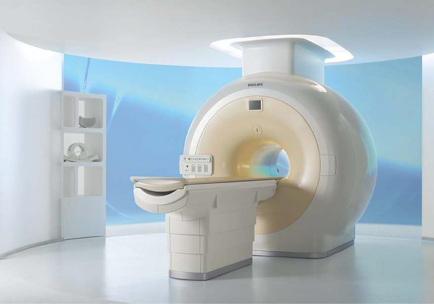
Equipment Description: Magnetic Resonance Imaging (MRI) is a well-established non-invasive technique for imaging the body and brain using strong magnetic fields and low energy radio waves to make pictures of the inside of the body. The MRI scanner allows for very detailed scans with the help of a magnetic field and pulses of radio wave energy. Anatomical scans, including High resolution T2-weighted, High resolution FLAIR (fluid attenuated), and Very High resolution T1-weighted scans were conducted on 554 participants. Functional scans, including Diffusion-weighted, Brain activation in resting state and Blood Perfusion when breathing 5% CO2 gas were taken on 147 participants.
Assessment Type: Centre or Home Assessment
Protocol
- Initial recruitment prioritised participants aged 65 and over, using a random sampling procedure to select eligible members of the health assessment sample.
- Later recruitment targeted those aged 50–64 years on a case-wise basis.
- Participants who provided PAXgene bloods and EDTA3 bloods during the health assessment and did not meet the exclusion criteria were provided with information on TILDAs MRI study.
- Participants were contacted 10 days following this to schedule appointments.
- Participants attend CAMI in St. James’s Hospital for the MRI.
- A TILDA employee met participants at the reception and brought them to the centre.
- Participants complete a safety questionnaire on the day of the scan with a radiographer to ensure their safety.
- Participants lie on a cushioned platform inside the MRI scanner and are given earplugs/ear defenders and a mask to wear during the assessment.
- The MRI scan lasts 45 minutes.
- Anatomical scans, including High resolution T2-weighted, High resolution FLAIR (fluid attenuated), and Very High resolution T1-weighted scans were conducted on 555 participants.
- Functional scans, including Diffusion-weighted, Brain activation in resting state and Blood Perfusion when breathing 5% CO2 gas were taken on 147 participants.
Exclusion criteria included
- Participant less than 65 years
- Participant has a stent
- Participant has a cardiac pacemaker or defibrillator
- Participant has an implanted neurostimulator
- Participant has an implanted drug delivery device
- Participant has aneurysm clips in the brain
- Participant has a cochlear implant
Collection Waves
| Measure | Wave 1 | Wave 2 | Wave 3 | Wave 4 | Wave 5 | Wave 6 |
|---|---|---|---|---|---|---|
| MRI | X |
MRI Sequences
| Sequence | Description |
|---|---|
| High-resolution 2D T2-weighted | High-resolution T2-weighted anatomical sequence. |
| High-resolution 3D T1-weighted volumetric (IR-SPGR) | Three-dimensional T1-weighted volumetric anatomical scan acquired using an inversion recovery spoiled gradient recalled (IR-SPGR) sequence. |
| Fluid Attenuated Inversion Recovery (FLAIR) | T2-weighted FLAIR sequence for the assessment of white matter hyperintensities and other structural abnormalities. |
| Diffusion Weighted Imaging (DWI) | Four b-value acquisition for apparent diffusion coefficient (ADC) quantification. |
| Diffusion Tensor Imaging (DTI) | 61 diffusion encoding directions with a b-value of 1200 s/mm². |
| Pseudo-Continuous Arterial Spin Labelling (pCASL) | Single-shot echo-planar imaging (EPI) sequence with background suppression for cerebral perfusion measurement. |
| Functional MRI (BOLD) | Blood Oxygenation Level Dependent (BOLD) functional MRI sequence for assessment of brain activity and functional connectivity. |
Citation
- Citation: Jovicich, J., Marizzoni, M., Sala-Llonch, R., Bosch, B., Bartrés-Faz, D., Arnold, J., Benninghoff, J., Wiltfang, J., Roccatagliata, L., Nobili, F. and Hensch, T. (2013). Brain morphometry reproducibility in multi-center 3 T MRI studies: a comparison of cross-sectional and longitudinal segmentations. Neuroimage, 83, pp.472-484.
Example TILDA Papers
Leidhin, C.N., McMorrow, J., Carey, D., Newman, L., Williamson, W., Fagan, A.J., Chappell, M.A., Kenny, R.A., Meaney, J.F. and Knight, S.P. (2021). Age-related normative changes in cerebral perfusion: Data from The Irish Longitudinal Study on Ageing (TILDA). Neuroimage, 229, p.117741.
Shirsath, M.A., O’Connor, J.D., Boyle, R., Newman, L., Knight, S.P., Hernandez, B., Whelan, R., Meaney, J.F. and Kenny, R.A. (2024). Slower speed of blood pressure recovery after standing is associated with accelerated brain ageing: Evidence from The irish longitudinal study on ageing (TILDA). Cerebral circulation-cognition and behavior, 6, p.100212.
Zúñiga, R.G., Davis, J.R., Boyle, R., De Looze, C., Meaney, J.F., Whelan, R., Kenny, R.A., Knight, S.P. and Ortuño, R.R. (2023). Brain connectivity in frailty: insights from The Irish Longitudinal Study on Ageing (TILDA). Neurobiology of aging, 124, pp.1-10.